变形体
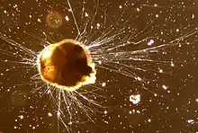
变形体(amoeba),[1]是一种通过伸长或收回伪足来改变自身形状的细胞或生物。[2]变形体并不属于某个单一的生物分类,相反地,在许多真核生物中都能找到它们的存在。变形体细胞不仅存在于原生动物中,也广泛存在于各种真菌、藻类以及动物中。[3][4][5][6][7]
微生物学家经常会使用术语变形体来称呼那些能施展变形运动的生物。[8][9]
在旧的分类系统中,大多数的变形体都被分在了肉足纲或肉足亚门(一类通过产生伪足而缓慢移动的单细胞生物)。然而,分子系统发生学研究已经表明肉足纲并不是有着共同起源的一类单系群。因此,变形体不再被定义为一类单一种群生物。[10]
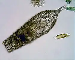
最著名的变形体原生生物巨型阿米巴属于卡罗莱纳卓变形虫和大变形虫,二者均已在实验室中被广泛培养或研究。[11][12]其他的著名种类包括所谓的食脑变形虫——福氏耐格里变形虫;肠道寄生的、引起阿米巴原虫病的溶组织内阿米巴;以及多细胞的一种黏菌--盘基网柄菌。
形状、运动与食物
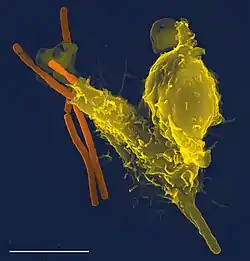
变形体使用伪足移动并进食。这些伪足是由凸起的细胞质经协同作用而形成的肌动蛋白与质膜细胞周围的微丝结合而成的。[13]
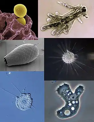
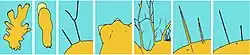
伪足的外观和内部结构可用于区分不同类型的变形体。变形虫门的物种, 如变形虫通常有球根状的伪足,其伪足拥有圆形的两端,横断面呈管状;而丝足虫门的物种,如鳞壳虫属与网足虫属的物种,则有着线一样的伪足;有孔虫门生物则发出细的并带有分支的伪足,大量的分支在合并到一起还可以形成网状结构。有些物种,如放射虫和太阳虫,这些生物的伪足拥有针状的伪足,并且十分坚硬,由内部的微管向外延伸而成。[3][14]
.png.webp)
变形体可分为两类:有壳的与无壳的。壳是由多种复杂的物质组成的,包括钙,二氧化硅,甲壳素或是类似硅藻粘合细胞壁所用的像沙子的小颗粒。[15]
为维持渗透压,大多数的淡水变形虫拥有可收缩的液泡将多余的水从细胞内挤走。[16] 这是因为在淡水中必须要有一个相比变形虫自己内部的流体较低的浓度溶质。因为周围的水相对于细胞内部处于低渗状态,水需要传送到变形虫的细胞膜进行渗透。如果没有液泡进行收缩的话,细胞就会充满大量多余的水分,最终会导致细胞破裂,死亡。
而海洋中的变形虫通常不具备可伸缩的液泡因为细胞内溶质浓度与周围的水大致保持平衡。[17]
变形体的食物来源通常是变化的。一些变形体会捕食周围的细菌或原生动物。而有些则是食腐者,它们会食用死亡生物的碎屑。
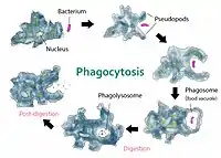
变形体通常通过吞噬作用来进食,它们在进食时伸出伪足,包围并吞噬它们的猎物。变形虫细胞没有嘴进行吞咽,因此它们并没有固定的地方进行吞噬作用。[18]
大小
变形体的细胞或变形体本身的大小经常是变化的。下图是一些变形体的大小。
| 细胞或物种名称 | 大小(微米) |
|---|---|
| 多聚覆毛虫[20] | 2.3–3 |
| 福氏耐格里变形虫[21] | 8–15 |
| 中性粒细胞(一种白细胞)[22] | 12–15 |
| 棘变形虫属[23] | 12–40 |
| 溶组织内阿米巴[24] | 15–60 |
| 蕈状变形虫[25] | 30–152 |
| 大变形虫[26] | 220–760 |
| 卡罗莱纳卓变形虫[27] | 700–2000 |
| 沼泽多核变形虫[28] | 最大能达到5000 |
| 脆性介壳虫[29] | 最大能达到200000 |
变形体的生命周期
一些多细胞生物只在某些特定的阶段才拥有变形体细胞,而某些生物变形体细胞只能用于某些特定的功能。人类和其他动物在免疫系统中,变形体白细胞只负责追捕入侵的微生物,如细菌和病原体,然后将它们吞噬。[30]
在多细胞真菌类生物中也有着变形体的存在,比如所谓的粘菌。无论是粘菌(目前分类为黏菌纲),以及产生混杂胶丝的细胞粘菌和网柱原虫目,都有着变形体阶段来进行摄食。前者结合形成一个巨大的变形体细胞多核生物,[31] 而后者的细胞分开利用,直到能源和食物耗尽,此时多个变形体则聚集成多为一个单一的机体。[8]
其他的生物也可以在特定的生命周期阶段产生变形体细胞,如在一些绿色藻类(双星藻纲)[32]和壳缝羽纹矽藻[33]产生配子的时候。
与其他生物的致病作用
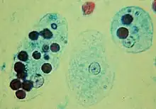
有些变形体可以感染其他生物致病性,导致疾病:
参考
- "Amoeba" (页面存档备份,存于) at Oxforddictionaries.com
- Singleton, Paul. . Chichester, UK: John Wiley & Sons. 2006: 32. ISBN 978-0-470-03545-0.
- David J. Patterson. . Tree of Life web project. [2018-04-12]. (原始内容存档于2010-06-15).
- . The University of Edinburgh. [2018-04-12]. (原始内容存档于2009-06-10).
- Wim van Egmond. . Microscopy-UK. [2018-04-12]. (原始内容存档于2005-11-04).
- Flor-Parra, Ignacio; Bernal, Manuel; Zhurinsky, Jacob; Daga, Rafael R. . Biology Open. 2013-12-17, 3 (1): 108–115. ISSN 2046-6390. PMC 3892166
 . PMID 24357230. doi:10.1242/bio.20136783.
 - Friedl, P.; Borgmann, S.; Bröcker, E. B. . Journal of Leukocyte Biology. 2001-10-01, 70 (4): 491–509. ISSN 0741-5400. PMID 11590185.
- Marée, Athanasius FM, and Paulien Hogeweg. "How amoeboids self-organize into a fruiting body: multicellular coordination in Dictyostelium discoideum." Proceedings of the National Academy of Sciences 98.7 (2001): 3879–3883.
- Mackerras, M. J., and Q. N. Ercole. "Observations on the action of paludrine on malarial parasites." Transactions of the Royal Society of Tropical Medicine and Hygiene 41.3 (1947): 365–376.
- Jan Pawlowski: The twilight of Sarcodina: a molecular perspective on the polyphyletic origin of amoeboid protists. Protistology, Band 5, 2008, S. 281–302. (pdf, 570 kB) (页面存档备份,存于)
- Tan; et al. (PDF). Protistology. 2005, 4: 185–90 [2018-04-12]. (原始内容存档 (PDF)于2017-09-29).
- . Amoeba proteus. 2013-04-12 [2017-09-28]. (原始内容存档于2017-09-29) (美国英语).
- Alberts Eds.; et al. . New York: Garland Science. 2007: 1037. ISBN 9780815341055.
- Margulis, Lynn. . Academic Press. 2009: 206–7. ISBN 978-0-12-373621-5.
- Ogden, C. G. . Oxford, London, Glasgow: Oxford University Press, for British Museum (Natural History). 1980: 1–5. ISBN 0198585020.
- Alberts Eds.; et al. . New York: Garland Science. 2007: 663. ISBN 9780815341055.
- Kudo, Richard Roksabro. "Protozoology." Protozoology 4th Edit (1954). p. 83
- Thorp, James H. (2001). Ecology and Classification of North American Freshwater Invertebrates. San Diego: Academic. p. 71. ISBN 0-12-690647-5.
- Jeon, Kwang W. . New York: Academic Press. 1973: 100.
- Mylnikov, Alexander P.; Weber, Felix; Jürgens, Klaus; Wylezich, Claudia. . European Journal of Protistology. 2015-08-01, 51 (4): 299–310. ISSN 1618-0429. PMID 26163290. doi:10.1016/j.ejop.2015.05.002.
- . [2016-08-21]. (原始内容存档于2016-08-21) (美国英语).
- . www.anatomyatlases.org. [2016-08-21]. (原始内容存档于2016-08-19).
- . www.arcella.nl. [2016-08-21]. (原始内容存档于2016-08-18).
- . msu.edu. [2016-08-21]. (原始内容存档于2016-10-05).
- . www.arcella.nl. [2016-08-21]. (原始内容存档于2016-08-18).
- . www.arcella.nl. [2016-08-21]. (原始内容存档于2016-08-18).
- . www.arcella.nl. [2016-08-21]. (原始内容存档于2016-10-12).
- . www.arcella.nl. [2016-08-21]. (原始内容存档于2016-08-18).
- Gooday, A. J.; Aranda da Silva, A.; Pawlowski, J. . Deep-Sea Research Part II: Topical Studies in Oceanography. The Geology, Geochemistry, and Biology of Submarine Canyons West of Portugal. 2011-12-01, 58 (23–24): 2401–2419 [2018-04-12]. doi:10.1016/j.dsr2.2011.04.005. (原始内容存档于2018-10-04).
- Friedl, Peter, Stefan Borgmann, and Eva-B. Bröcker. "Amoeboid leukocyte crawling through extracellular matrix: lessons from the Dictyostelium paradigm of cell movement." Journal of Leukocyte Biology 70.4 (2001): 491–509.
- Nakagaki; et al. . Nature. 2000, 407 (6803): 470 [14 September 2014]. PMID 11028990. doi:10.1038/35035159. (原始内容存档于2017-06-26).
- Wehr, John D. . San Diego and London: Academic Press. 2003: 353. ISBN 978-0-12-741550-5.
- . Algae World. Royal Botanic Garden Edinburgh. [1 March 2015]. (原始内容存档于2014-09-23).
外部链接
| 維基文庫在1905版《新國際百科全書》有Rhizopoda的描述。(英文) |
| 维基共享资源上的相关多媒体资源:变形体 |
- Siemensma, F. Microworld: world of amoeboid organisms (页面存档备份,存于).
- Völcker, E. & Clauß, S. Visual key to amoeboid morphotypes (页面存档备份,存于). Penard Labs.
- The Amoebae website of Maciver Lab of the University of Edinburgh, brings together information from published sources.
- Molecular Expressions Digital Video Gallery: Pond Life – Amoeba (Protozoa) (页面存档备份,存于) – informative amoeba videos