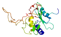
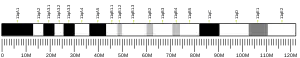
SOCS3
细胞因子信号传导抑制因子3(SOCS3或 SOCS-3)是一种在人体内由 SOCS3基因编码的蛋白。[5][6] 该基因编码 STAT- 诱导的 STAT 抑制子(SSI),也可称为细胞因子信号转导(SOCS)家族抑制子,的一个成员。 SSI 家族成员是可由细胞因子诱导的细胞因子信号转导负调节因子。
功能
SOCS3基因的表达可由多种细胞因子的诱导,包括 IL-6,IL-10,干扰素-γ(IFN-γ)。
在 IL-6,Epo,GCSF 和瘦素的信号传导中,已发现SOCS3在各自的细胞因子受体上的结合对 SOCS3的抑制功能而言是至关重要的。
SOCS3的过表达会抑制脂肪组织和肝脏胰岛素信号转导,但对肌肉则不然。[7] 但是在小鼠骨骼肌中敲除 SOCS3可以保护小鼠免于肥胖相关的胰岛素抵抗。[7]
由于神经酰胺合成的增加,SOCS3会增加瘦素抵抗和胰岛素抵抗。[8] 因此,研究已表明去除 SOCS 基因可以预防肥胖症患者的胰岛素抵抗[7]
对该小鼠中基因的同源基因的研究表明,该基因在胎儿肝脏造血和胎盘发育的负调控中发挥作用。[9]
SOCS3蛋白能与JAK2结合,抑制其活性。
相互作用
已经证明 SOCS3可以与下列物质相互作用:
参见
- SOCS
- JAK-STAT信号通路
参考文献
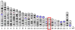
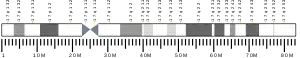
- GRCh38: Ensembl release 89: ENSG00000184557 - Ensembl, May 2017
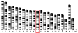
- GRCm38: Ensembl release 89: ENSMUSG00000053113 - Ensembl, May 2017
- . National Center for Biotechnology Information, U.S. National Library of Medicine.
- . National Center for Biotechnology Information, U.S. National Library of Medicine.
- . Biochem Biophys Res Commun. September 1997, 237 (1): 79–83. PMID 9266833. doi:10.1006/bbrc.1997.7080.
- . Biochem Biophys Res Commun. November 1997, 239 (2): 439–46. PMID 9344848. doi:10.1006/bbrc.1997.7484.
- . Diabetes. 2013, 62 (1): 56–64 [2018-11-22]. PMC 3526029
 . PMID 22961088. doi:10.2337/db12-0443. (原始内容存档于2018-11-23).
 - . American Journal of Physiology. 2009, 297 (1): E211–E224 [2018-11-22]. PMC 2711669 . PMID 19435851. doi:10.1152/ajpendo.91014.2008. (原始内容存档于2016-08-10).
- . (原始内容存档于2010-03-07).
- . J. Biol. Chem. September 2000, 275 (38): 29338–47. PMID 10882725. doi:10.1074/jbc.M003456200.
- . Eur. J. Biochem. May 2002, 269 (10): 2516–26. PMID 12027890. doi:10.1046/j.1432-1033.2002.02916.x.
- . J. Biol. Chem. January 2003, 278 (1): 661–71. PMID 12403768. doi:10.1074/jbc.M210552200.
- . Biochem. Biophys. Res. Commun. November 2000, 278 (1): 38–43. PMID 11071852. doi:10.1006/bbrc.2000.3762.
- . Genes Cells. June 1999, 4 (6): 339–51. PMID 10421843. doi:10.1046/j.1365-2443.1999.00263.x.
- . Nat. Cell Biol. May 2001, 3 (5): 460–5. PMID 11331873. doi:10.1038/35074525.
- . Cell Death Differ. 2008, 15 (7): 1187–95. PMID 18483491. doi:10.1038/cdd.2008.69.
- . Exp Dermatol. 2010, 19 (9): 854–6. PMID 20698882. doi:10.1111/j.1600-0625.2010.01118.x.
扩展阅读
- Yasukawa H, Sasaki A, Yoshimura A (2000). "Negative regulation of cytokine signaling pathways". Annu. Rev. Immunol. 18: 143–64. doi:10.1146/annurev.immunol.18.1.143. PMID 10837055(页面存档备份,存于).
- Kile BT, Schulman BA, Alexander WS, Nicola NA, Martin HM, Hilton DJ (2002). "The SOCS box: a tale of destruction and degradation". Trends Biochem. Sci. 27 (5): 235–41. doi:10.1016/S0968-0004(02)02085-6. PMID 1276535(页面存档备份,存于).
- Zhang JG, Farley A, Nicholson SE, Willson TA, Zugaro LM, Simpson RJ, Moritz RL, Cary D, Richardson R, Hausmann G, Kile BJ, Kent SB, Alexander WS, Metcalf D, Hilton DJ, Nicola NA, Baca M (1999). "The conserved SOCS box motif in suppressors of cytokine signaling binds to elongins B and C and may couple bound proteins to proteasomal degradation". Proc. Natl. Acad. Sci. U.S.A. 96 (5): 2071–6. doi:10.1073/pnas.96.5.2071. PMC 26738. PMID 10051596(页面存档备份,存于).
- Sasaki A, Yasukawa H, Suzuki A, Kamizono S, Syoda T, Kinjyo I, Sasaki M, Johnston JA, Yoshimura A (1999). "Cytokine-inducible SH2 protein-3 (CIS3/SOCS3) inhibits Janus tyrosine kinase by binding through the N-terminal kinase inhibitory region as well as SH2 domain". Genes Cells. 4 (6): 339–51. doi:10.1046/j.1365-2443.1999.00263.x. PMID 10421843(页面存档备份,存于).
- Marine JC, McKay C, Wang D, Topham DJ, Parganas E, Nakajima H, Pendeville H, Yasukawa H, Sasaki A, Yoshimura A, Ihle JN (1999). "SOCS3 is essential in the regulation of fetal liver erythropoiesis". Cell. 98 (5): 617–27. doi:10.1016/S0092-8674(00)80049-5. PMID 10490101(页面存档备份,存于).
- Schmitz J, Weissenbach M, Haan S, Heinrich PC, Schaper F (2000). "SOCS3 exerts its inhibitory function on interleukin-6 signal transduction through the SHP2 recruitment site of gp130". J. Biol. Chem. 275 (17): 12848–56. doi:10.1074/jbc.275.17.12848. PMID 10777583(页面存档备份,存于).
- Ram PA, Waxman DJ (2000). "SOCS/CIS protein inhibition of growth hormone-stimulated STAT5 signaling by multiple mechanisms". J. Biol. Chem. 274 (50): 35553–61. doi:10.1074/jbc.274.50.35553. PMID 10585430(页面存档备份,存于).
- Sasaki A, Yasukawa H, Shouda T, Kitamura T, Dikic I, Yoshimura A (2000). "CIS3/SOCS-3 suppresses erythropoietin (EPO) signaling by binding the EPO receptor and JAK2". J. Biol. Chem. 275 (38): 29338–47. doi:10.1074/jbc.M003456200. PMID 10882725(页面存档备份,存于).
- Anhuf D, Weissenbach M, Schmitz J, Sobota R, Hermanns HM, Radtke S, Linnemann S, Behrmann I, Heinrich PC, Schaper F (2000). "Signal transduction of IL-6, leukemia-inhibitory factor, and oncostatin M: structural receptor requirements for signal attenuation". J. Immunol. 165 (5): 2535–43. doi:10.4049/jimmunol.165.5.2535. PMID 10946280(页面存档备份,存于).
- Bjorbak C, Lavery HJ, Bates SH, Olson RK, Davis SM, Flier JS, Myers MG (2001). "SOCS3 mediates feedback inhibition of the leptin receptor via Tyr985". J. Biol. Chem. 275 (51): 40649–57. doi:10.1074/jbc.M007577200. PMID 11018044(页面存档备份,存于).
- Shen X, Hong F, Nguyen VA, Gao B (2000). "IL-10 attenuates IFN-alpha-activated STAT1 in the liver: involvement of SOCS2 and SOCS3". FEBS Lett. 480 (2–3): 132–6. doi:10.1016/S0014-5793(00)01905-0. PMID 11034314(页面存档备份,存于).
- Dey BR, Furlanetto RW, Nissley P (2001). "Suppressor of cytokine signaling (SOCS)-3 protein interacts with the insulin-like growth factor-I receptor". Biochem. Biophys. Res. Commun. 278 (1): 38–43. doi:10.1006/bbrc.2000.3762. PMID 11071852(页面存档备份,存于).
- Hartley JL, Temple GF, Brasch MA (2001). "DNA cloning using in vitro site-specific recombination" (页面存档备份,存于). Genome Res. 10 (11): 1788–95. doi:10.1101/gr.143000. PMC 310948 (页面存档备份,存于). PMID 11076863(页面存档备份,存于).
- Cookson WO, Ubhi B, Lawrence R, Abecasis GR, Walley AJ, Cox HE, Coleman R, Leaves NI, Trembath RC, Moffatt MF, Harper JI (2001). "Genetic linkage of childhood atopic dermatitis to psoriasis susceptibility loci". Nat. Genet. 27 (4): 372–3. doi:10.1038/86867. PMID 11279517(页面存档备份,存于).
- Cacalano NA, Sanden D, Johnston JA (2001). "Tyrosine-phosphorylated SOCS-3 inhibits STAT activation but binds to p120 RasGAP and activates Ras". Nat. Cell Biol. 3 (5): 460–5. doi:10.1038/35074525. PMID 11331873(页面存档备份,存于).
- Roberts AW, Robb L, Rakar S, Hartley L, Cluse L, Nicola NA, Metcalf D, Hilton DJ, Alexander WS (2001). "Placental defects and embryonic lethality in mice lacking suppressor of cytokine signaling 3". Proc. Natl. Acad. Sci. U.S.A. 98 (16): 9324–9. doi:10.1073/pnas.161271798. PMC 55419. PMID 11481489(页面存档备份,存于).
- Dif F, Saunier E, Demeneix B, Kelly PA, Edery M (2001). "Cytokine-inducible SH2-containing protein suppresses PRL signaling by binding the PRL receptor". Endocrinology. 142 (12): 5286–93. doi:10.1210/endo.142.12.8549. PMIDPMID 11713228(页面存档备份,存于).
- Bode JG, Ludwig S, Freitas CA, Schaper F, Ruhl M, Melmed S, Heinrich PC, Häussinger D (2002). "The MKK6/p38 mitogen-activated protein kinase pathway is capable of inducing SOCS3 gene expression and inhibits IL-6-induced transcription". Biol. Chem. 382 (10): 1447–53. doi:10.1515/BC.2001.178. PMID 11727828(页面存档备份,存于).
This article is issued from Wikipedia. The text is licensed under Creative Commons - Attribution - Sharealike. Additional terms may apply for the media files.